
EKG Interpretation for Nurses: A Step-by-Step Guide
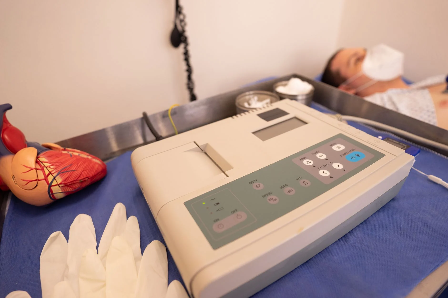
Accurate ekg interpretation for nurses is a foundational skill for patient safety in telemetry, critical care, and general medical-surgical units. While automated monitor readings provide a starting point, they can be misled by artifact or patient movement. A systematic approach ensures you identify life-threatening arrhythmias independently and correlate findings with the patient’s clinical status. This guide provides a repeatable framework for analyzing cardiac rhythms. Always remember that your institutional policy dictates your specific scope of practice and escalation protocols, as recommended by the American Association of Critical-Care Nurses (AACN) standards for patient-centered monitoring.
The 6-Step Systematic Approach
To ensure consistency and safety, follow this repeatable checklist for every rhythm strip. This method minimizes diagnostic errors and helps you identify basic ekg rhythms effectively.
- Rate: Determine the atrial and ventricular rate. For regular rhythms, count the number of R-waves in a 6-second strip and multiply by 10. For precise measurement, count small boxes between R-R intervals (1500 divided by the number of small boxes).
- Rhythm: Assess regularity. Use calipers or a piece of paper to mark R-R intervals. If the distance between R-waves varies, the rhythm is irregular.
- P-waves: Identify P-waves. Are they present, upright, and uniform? Is there one P-wave before every QRS complex?
- PR Interval: Measure from the start of the P-wave to the beginning of the QRS complex. While common references cite 0.12 to 0.20 seconds, always verify the specific threshold defined by your institutional protocol.
- QRS Duration: Measure the width of the QRS complex. While common references cite 0.06 to 0.10 seconds, interpret this width in the context of your patient’s baseline and facility guidelines.
- QT Interval: Measure from the start of the QRS to the end of the T-wave. This interval is rate-dependent; use your facility’s standard QTc calculation tool to assess for potential risks.
Normal EKG Intervals and Values
Use this table as a quick reference for cardiac electrical activity. Note that these values are educational baselines; your institutional policy may have specific parameters for clinical escalation.
| Measurement | Clinical Reference |
|---|---|
| Heart Rate | 60–100 bpm (varies by patient baseline) |
| PR Interval | 0.12–0.20 seconds (typical range) |
| QRS Duration | 0.06–0.10 seconds (typical range) |

Clinical Escalation: When to Notify the Provider
Your role in monitoring involves prioritizing patient-centered assessment over machine-generated alerts. If you observe a rhythm that deviates from the patient’s baseline, assess the patient immediately. Check for a pulse, blood pressure, and level of consciousness. If the patient is symptomatic, follow your facility’s rapid response or ACLS protocols. Notify the provider immediately for new-onset ventricular arrhythmias, high-grade heart blocks, sustained tachycardia or bradycardia with hemodynamic instability, or significant changes in the ST segment. Always document your findings and the patient’s clinical response in the electronic health record according to your unit’s policy. If you are looking to expand your knowledge of related cardiac care, consider reviewing resources on pharmacology for nurses to understand how common medications influence cardiac rhythms.
Rhythm Strip vs. 12-Lead EKG
Nurses often work with both rhythm strips and 12-lead EKGs. A rhythm strip, usually Lead II, provides a continuous view of the heart’s electrical activity, which is essential for monitoring rate and rhythm changes over time. In contrast, a 12-lead EKG provides a 3-dimensional view of the heart’s electrical activity from 12 different angles. Use the 12-lead EKG when you need to identify the location of an infarction, evaluate bundle branch blocks, or investigate complex arrhythmias. While a rhythm strip is your primary tool for ongoing monitoring, the 12-lead is a diagnostic snapshot.
Legal Scope and Documentation
The nurse’s legal scope of practice regarding cardiac monitoring is defined by individual state boards of nursing and specific institutional policies. While nurses are expected to identify and report lethal arrhythmias, the final medical diagnosis remains the responsibility of the provider. Documentation must be objective, noting the rhythm observed, the patient’s clinical presentation, and any interventions performed. For those involved in specialized care, maintaining current knowledge of legal and ethical standards is vital, which you can explore through ethics CE for nurses.
Differentiating Between Artifact and True Arrhythmia
One of the most common challenges in ekg interpretation for nurses is distinguishing between a life-threatening arrhythmia and signal artifact. Artifact—often caused by loose electrodes, patient movement, muscle tremors, or electromagnetic interference—can mimic ventricular tachycardia or fibrillation on a monitor. Before initiating a code or rapid response, always perform a visual check of the patient. If the rhythm on the monitor appears lethal but the patient is alert, talking, and hemodynamically stable, the rhythm is likely artifact.
To troubleshoot, ensure all leads are securely attached and that the skin is clean and dry. If the rhythm persists, check the patient’s pulse manually at a peripheral site. If a strong, regular pulse is present despite a chaotic monitor reading, prioritize the physical assessment over the screen. Always document the clinical correlation; for example, note that the patient was “asymptomatic with a palpable pulse despite monitor artifact.”
Calculating Heart Rate on Irregular Rhythms
Standard rate calculation methods like the “1500 method” or “300-150-100” rule rely on a regular R-R interval. When dealing with irregular rhythms such as atrial fibrillation, these methods will provide inaccurate results. Instead, use the 6-second strip method to determine the ventricular rate.
Most telemetry paper is marked with hash marks at the top or bottom of the strip, representing 3-second intervals. Count the number of R-waves in two of these 3-second segments (a 6-second window) and multiply that number by 10. This provides an estimate of the beats per minute. For a more precise calculation over a longer duration, count the R-waves in a 1-minute strip. Many nurses find it useful to keep a small, laminated ekg interpretation pdf at the workstation, which includes a visual guide for calculating rates on irregular strips, ensuring consistency across shifts.
Basic EKG Rhythms Reference Guide
Recognizing basic ekg rhythms requires a systematic evaluation of the P-QRS-T relationship. Use this table to differentiate common rhythm patterns based on their electrical characteristics.
| Rhythm | Rate (bpm) | P-Waves | QRS Duration | Regularity |
|---|---|---|---|---|
| Normal Sinus | 60–100 | Present, uniform | < 0.12s | Regular |
| Sinus Bradycardia | < 60 | Present, uniform | < 0.12s | Regular |
| Sinus Tachycardia | > 100 | Present, uniform | < 0.12s | Regular |
| Atrial Fibrillation | Variable | Absent (fibrillatory waves) | < 0.12s | Irregularly irregular |
| Ventricular Tachycardia | > 100 | Absent | > 0.12s (wide) | Usually regular |
The Role of the Nurse in EKG Monitoring
Your role extends beyond identifying rhythms; you are the frontline for clinical decision-making. Effective cardiac monitoring requires you to integrate rhythm data with the patient’s clinical status, medication profile, and electrolyte levels. For instance, hypokalemia or hypomagnesemia can significantly alter the QT interval, increasing the risk of Torsades de Pointes. Always verify that your monitoring parameters, such as alarm limits for heart rate and ST-segment changes, are individualized to the patient’s baseline per unit policy.
When an alarm sounds, your priority is to assess the patient first, then the monitor. If you identify a new-onset arrhythmia, gather objective data—including the patient’s blood pressure, oxygen saturation, and mental status—before notifying the provider. Clear, concise communication using the SBAR (Situation, Background, Assessment, Recommendation) format ensures that the provider has the necessary information to make an informed decision regarding the patient’s care.
Next Steps for Clinical Proficiency
Mastering EKG interpretation takes consistent practice. Many nurses find it helpful to keep an ekg interpretation pdf or reference card at the bedside for quick verification. If you are working toward a specialty certification such as the CCRN, ensure your study materials align with current AHA guidelines for rhythm management. Continue to refine your assessment skills through ongoing education and by participating in unit-based case reviews.
Frequently Asked Questions
What is the most important step in EKG interpretation?
The most important step is assessing the patient. Never rely solely on the monitor. If the monitor shows a lethal rhythm, immediately check the patient for a pulse and signs of perfusion. Clinical assessment always takes precedence over the rhythm strip.
How do I differentiate between artifact and a true arrhythmia?
Artifact is often caused by patient movement, loose electrodes, or electrical interference. If the rhythm looks abnormal, check the patient's lead placement and ask the patient to remain still. If the rhythm persists and the patient remains asymptomatic, verify the findings with a 12-lead EKG or a manual pulse check.
What should I do if the monitor shows a lethal rhythm but the patient is alert?
If the patient is alert and talking, the monitor reading is likely an artifact. Check the leads, ensure the patient is stable, and verify the heart rate manually by palpating a pulse. Do not initiate emergency protocols for an alert patient based on a suspicious monitor reading without confirming the cardiac status.
What are the common causes of EKG artifact?
Common causes of EKG artifact include patient movement (somatic tremor), loose or poorly adhered electrodes, dried-out gel on electrodes, electromagnetic interference from nearby electronic equipment, and cable movement.
Are there specific certifications for EKG interpretation?
While there is no single 'EKG interpretation certification,' nurses can validate their competency through specialty certifications like the CCRN or CEN, which include cardiac rhythm mastery as a core component. Always check with your facility regarding internal competency validation requirements.
About This Article
Written by the Nurse CE Hub Editorial Team. We base state requirements and certification details on official board of nursing and certifying-organization sources, linked within the article.
Rules and fees can change. Confirm current requirements with your state board of nursing or certifying organization before you renew or apply. This article is general information, not legal or professional advice. How we research our content